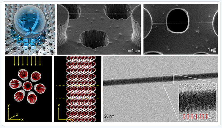
电子显微镜下的DNA双螺旋结构
时间: 2012-12-04 点击次数:次 作者:admin
Direct Imaging of DNA Fibers: The Visage of Double Helix
Francesco Gentile, Manola Moretti, Tania Limongi Andrea Falqui, Giovanni Bertoni, Alice Scarpellini, Stefania Santoriello, Luca Maragliano,Remo Proietti Zaccaria, and Enzo di Fabrizio
Nanostructures, Neuroscience and Brain Technologies, and Nanochemistry Departments, Istituto Italiano diTecnologia, Via Morego 30, 16163 Genova, Italy
BIONEM, Bio-Nanotechnology and Engineering for Medicine, Department of experimental and clinical medicine, University of Magna Graecia Viale Europa, Germaneto, 88100 Catanzaro, Italy
IMEM-CNR, Parco Area delle Scienze 37/A, 43124 Parma, Italy
Nano Lett., Article ASAP DOI:10.1021/nl3039162
Publication Date (Web): November 22, 2012
Copyright ?2012 American Chemical Society
*E-mail:enzo.difabrizio@iit.it.

Abstract
Direct imaging becomes important when the knowledge at few/single molecule level is requested and where the diffraction does not allow to get structural and functional information. Here we report on the direct imaging of double stranded (ds) ?DNA in the A conformation, obtained by combining a novel sample preparation method based on super hydrophobic DNA molecules self-aggregation process with transmission electron microscopy (TEM). The experimental breakthrough is the production of robust and highly ordered paired DNA nanofibers that allowed its direct TEM imaging and the double helix structure revealing.
Keywords:
DNA bundles direct imaging;transmission electron microscopy;superhydrophobic surface;nanofabricated micropillars;molecular dynamics simulations
